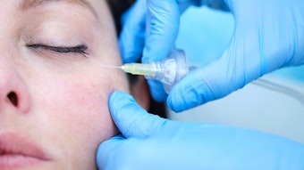
In an effort to reduce the risk of complications following glabellar soft tissue filler injections, researchers Sebastian Cotofana, MD, PhD, et al, used ultrasound imaging to identify the course of the superficial branch of the supratrochlear artery and of the deep branch of the supraorbital artery in relation to the ipsilateral vertical glabellar line. They measured the diameters, distance from skin surface, distance between the midline, distance between vertical glabella lines and the cutaneous projection of the supratrochlear/supraorbital arteries at rest and upon frowning.
After analyzing 41 healthy volunteers (mean age of 26.17 years and mean BMI of 23.09 kg/m2), they found that the mean depth of the supratrochlear artery was 3.34mm at rest and the mean depth of the supraorbital artery was 3.54mm.
The location of the arteries varied by gender: The mean distance between the superficial branch of the supratrochlear artery and the ipsilateral vertical glabellar line was 10.59mm in males and 8.21mm in females. The mean distance between the supraorbital artery and the ipsilateral vertical glabellar line was 22.38mm for in males and 20.73mm in females.
Upon frowning, there was a medial shift in the position of the supratrochlear arterial of 1.63mm in males and 1.84mm in females and a shift of 3.9mm in supraorbital arterial position for both genders .
The authors of the study, published in the Aesthetic Surgery Journal (December 2020), concluded, “The hypothesis that injecting soft tissue fillers next to the vertical glabellar line is safe because the supratrochlear artery courses deep to the crease should be rejected. Additionally, the glabella and the supraorbital region should be considered as an area of mobile, rather than static, soft tissues.”


