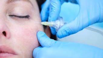
Mariana Calomeni, MD, et al, used ultrasound to evaluate the location and function of the angular vein in the tear trough in three different facial expressions (repose, smiling, and maximum orbicularis oculi contraction) to help improve the safety and efficacy of soft tissue filler injections in the tear trough.
Related: [Filler Safety] Understanding Orbital Arterial Distribution
The study, published in the Aesthetic Surgery Journal (May 2022), included 20 participants with a mean age of 48.3 years and mean BMI of 24.5 kg/m2. The diameter of the angular vein and the velocity and direction of venous blood flow were analyzed during the three facial expressions.
In 100% of the subjects, the angular vein traveled inside the orbicularis oculi muscle (intra-muscular course) within the tear trough. The angular artery was not identified in this location. The distance between the angular vein and the inferior orbital rim was (lateral to medial): 4.6 mm, 4.5 mm, 3.9 mm and 3.8 mm. The caudally directed blood flow was 10.2 cm/s in repose and 7.3 cm/s at maximum orbicularis oculi muscle contraction. No blood flow was detectable during smiling.
Related: [Filler Safety] Rethinking Glabella Injection Points
The authors concluded that “the diameter and the venous blood flow of the angular vein varied between the three tested facial expressions. Based on these anatomical findings, the deep injection approach to the tear trough is recommended due to the intramuscular course of the angular vein.”


