A study published in Plastic and Reconstructuve Surgery (March 2017) examined bacterial biofilm as a possible cause of adverse nodular reactions following treatment with filler injections. Researchers Mayuran Saththianathan, et al, performed an in vitro analysis of the propensity for three classes of filler materials to support biofilm growth; studied the effect of inoculation of filler material through a contaminated surface; and analyzed biopsy samples from patients presenting with chronic granulomatous inflammation following filler administration. Three fillers were included: hyaluronic acid (HA), polyacrylamide gel and poly-L-lactic acid.
To assess the fillers’ ability to promote biofilm formation in the presence/absence of additional nutrition, Staphylococcus epidermidis was diluted to 104 colony-forming units/ml in phosphate-buffered saline for the no-nutrition group (control), and in 100% tryptic soy broth for the nutrition group. Aliquots of each filler or control were deposited onto a glass coupon with collagen framework and placed onto six-well tissue culture plates. Each filler was inoculated with 0.1ml of culture containing 103 colony-forming units/ml of S. epidermidis and, after incubation, wells were sonicated and the number of bacteria infecting each filler was determined. Each condition was repeated 20 times. Additional samples were stained and examined by confocal laser scanning microscopy; the presence of biofilm was confirmed visually.
All three filler materials supported the growth of S. epidermidis in the presence and absence of tryptic soy broth. In the absence of nutrition, bacterial growth was significantly greater on the HA filler sample compared with the other fillers and control. Bacterial growth on poly-L-lactic acid filler samples was greater than that of polyacrylamide gel and control, but bacterial growth on the polyacrylamide gel filler samples was not different from the control substrate. In the presence of nutrition, polyacrylamide gel supported significantly more growth than either poly-L-lactic acid or HA, and the HA filler had significantly more growth than poly-L-lactic acid.
For the needle pass examination, researchers added 3ml of sloppy nutrient agar (0.5%) to the wells. They placed the open plates inside a sterile, tightly secured surgical glove. The glove area covering each well was inoculated with 100µl of tryptic soy broth and allowed to dry. A total of 1ml of HA filler was injected through the inoculated glove area. Each condition was repeated 17 times. There were10,000-fold higher numbers of bacteria infecting the filler with multiple passes compared with a single-pass injection of filler.
Biopsy results and microbiome analysis revealed multispecies biofilm, with an overall predominance of Pseudomonas and other intraoral and skin flora species. Common organisms forming 5% or more of the biofilm included Streptococcus mitis, S. salivarius, Staphylococcus aureus, coagulase-negative Staphylococci, Acidovorax species, Herbaspirillum putae, Ralstonia pickettii and Acinetobacter. The majority of bacteria were aerobic or facultative aerobic, with less than 10% of the microbiome composed of anaerobic organisms.
The researchers concluded that the study supports their hypothesis that biofilm is the likely cause of chronic granulomatous inflammation following filler injection, and they highlight the need for ongoing research to identify risk factors and techniques to prevent contamination at the time of injection.
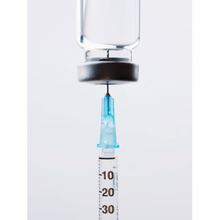
Photo copyright Getty Images.
